详情
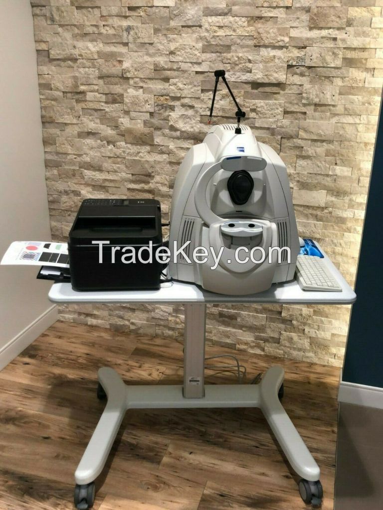
Employing the advanced imaging technology of spectral domain
optical coherence tomography, Cirrus HD-OCT acquires OCT data about
*0 times faster (*7,**0 vs. **0 A-scans per second) and with better
resolution (5 μm vs. ~*0 μm axial resolution in tissue), compared
to first-generation OCT technology. Cirrus acquires whole cubes of
OCT image data, composed of hundreds of line scans, in about the
same time as Stratus acquires a six-line scan. You can view these
data cubes in three planes, or through three dimensions, giving you
access to an extensive amount of retinal image data in one
scan.
Intended Use
The Cirrus HD-OCT with Retinal Nerve Fiber Layer (RNFL), Macular,
Optic Nerve Head, and Ganglion Cell Normative Databases is
indicated for in-vivo viewing, axial cross-sectional, and
three-dimensional imaging and measurement of anterior and posterior
ocular structures.
Indications for Use
The Cirrus HD-OCT is a non-contact, high resolution tomographic and
biomicroscopic imaging device. It is indicated for in-vivo viewing,
axial cross-sectional, and three-dimensional imaging and
measurement of anterior and posterior ocular structures, including
cornea, retina, retinal nerve fiber layer, ganglion cell plus inner
plexiform layer, macula, and optic nerve head. The Cirrus normative
databases are quantitative tools for the comparison of retinal
nerve fiber layer thickness, macular thickness, ganglion cell plus
inner plexiform layer thickness, and optic nerve head measurements
to a database of normal subjects. The Cirrus HD-OCT is intended for
use as a diagnostic device to aid in the detection and management
of ocular diseases including, but not limited to, macular holes,
cystoid macular edema, diabetic retinopathy, age-related macular
degeneration, and glaucoma.
Note: The Cirrus HD-OCT is not intended to be used as the sole
diagnostic for disease.
Essential Performance
The Essential Performance of the instrument is to provide accurate
measurements of anterior and posterior ocular structure.
Patient Population
The Cirrus HD-OCT may be used on all adults in need of diagnostic
evaluation of the eye. This includes (but is not limited to)
patients with the following disabilities or challenges:
• Wheelchair user
• Very low or not measurable visual acuity
• Fixation problems
• Postural problems
• Deafness
• Large body, but not those above *9th percentile based on
anthropomorphic data
There is a general requirement that the patient be able to sit
upright and be able to place their face in the chin and forehead
rest of the instrument (with or without supplemental human or
mechanical support).
Cirrus HD-OCT is designed for in-vivo viewing, axial
cross-sectional, and three-dimensional imaging and measurement of
anterior and posterior ocular structures.
Specifications:
HD-OCT Imaging
Methodology: Spectral Domain OCT
Optical Source: superluminescent diode (SLD), **0 nm
Optical Power: < **5 μW at the cornea
Scan Speed: *7,**0 A-scans per second
A-Scan depth: 2.0 mm (in tissue), ***4 points
Axial resolution: 5 μm (in tissue)
Transverse resolution: *5 μm (in tissue)
Fundus Imaging
Methodology: Line scanning ophthalmoscope
Live Fundus Image: During alignment and during OCT scan
Optical Source: Superluminescent diode (SLD), **0 nm
Optical Power: < 1.5 mW at the cornea
Field of View: *6 degrees W x *0 degrees H
Frame rate: >*0 Hz
Transverse resolution: *5 μm (in tissue)
Iris Imaging
Methodology: CCD Camera
Resolution: ***0 x ***4
Live iris image: During alignment
Electrical, Physical and Environemental
Weight: *8kg (*3 lbs)
Dimensions: *5L x *4W x *3H (cm)
Fixation: Internal, external
Internal fixation focus adjustment: **0D to **0D (diopters)
Input devices: Keyboard, mouse
Electrical rating (**5V) Single Phase, **0/**0V~ systems:****0Hz,
5A
Fuse rating (**5V) T 5A **0V
Electrical rating (**0V) Single Phase, **0/**0V~ systems:****0 Hz,
2.5A
Fuse rating (**0V) Fuse rating: T 5A **0V
Convenience Receptacle output ratings **5V~, 0.5 A Max, ****0
Hz
Temperature (transport and storage) **0º to **0º C
Relative humidity (transport and storage) *0% to **0%, including
condensation
Atmospheric pressure (transport and storage): **0 hPa to ***0
hPa
Temperature (operation) **0º to **5º C
Relative humidity (operation) *0% to *5%, excluding
condensation
Atmospheric Pressure (operation) **0 hPa to ***0 hPa
Computer
• High performance multi-core processor
• Internal storage: > *0,**0 scans
• CD-RW, DVD-ROM drive
• Integrated *5" color flat panel display