看起來你不是TradeKey.com的會員。 立即註冊,與全球超過7百萬的進口商和出口商建立聯繫。 点击注册--免费! |
BOOK A CALL
Book Call On Your Favorite Time
Code
🗘

By Signing Up. I agree to TradeKey.com Terms of Use, Privacy Policy, IPR and receive emails related to our services

Contact Us
product
Prev
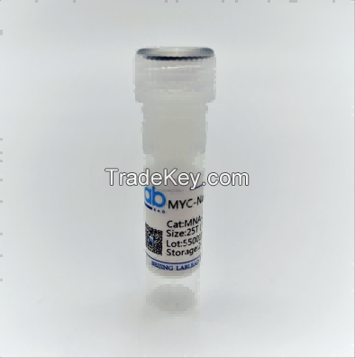
MYC-Nanoab-Agarose beads
Next

MYC-Nanoab-Agarose beads

离岸价格

获取最新报价

|

- Minimum Order

国家:

China

型号:

-

离岸价格:

获取最新报价

位置:

China

最小订单价格:

-

最小订单:

-

包装细节:

-

交货时间:

7-15 days

供应能力:

-

付款方式:

T/T, L/C, D/A, D/P, Western Union, Money Gram, PayPal, Other

產品組 :

现在联系
1st 年

联系人 Ying

现在联系

产品规格

详情

MYC-Nanoab-Agarose beads CATMNA*******0 Price: $**0 for *0T(negotiable)
product ID:GNA
Storage conditions:Store at **0°C, MYC-Agarose beads for regular use can be stored at 4 °C, and long-term storage at **0 or **0°C. Avoid centrifugation, drying and freeze-thaw.
 
Product description
Agarose beads conjugated with anti-MYC-tag nanoantibody were used for immunoprecipitation of MYC-tag ( MYC) fusion protein.
Product advantages
No heavy chain and light chain, clean background;
Ready-to use type, saving time;
High affinity and high binding ability.
Scope of application
It can be used for immunoprecipipation(IP)/immunocoprecipipation(CoIP)、chromatin immunoprecipitation(ChIP)/RNA-binding protein immunoprecipipation(RIP), enzyme activity determination, mass spectrometry analysis, etc.
Specificity
The specific binding is located at the N-terminal, C-terminal and internal MYC-tag of the fusion protein.
Product features
Storage buffer: PBS(containing *0%  ethanol);
Storage conditions: Stored at 4℃ for one year(to ensure full sealing), avoid centrifugation, drying and freeze-thaw.
 
Experimental steps:
Plant tissue cracking treatment:
Take an appropriate amount of plant tissue samples (leaves, etc.) and place them in frozen liquid nitrogen, and then put the frozen plant tissue samples in a mortar for grinding, so as to fully grind and destroy their cell walls. Add *******0ul RIPA lysate (added with protease inhibitor PMSF, etc.) for cracking. In order to improve the cracking efficiency, add **0ul glass powder and fully shake for *0min. After cracking, ****0 rpm, centrifuge for *0min, suck the supernatant and place it in a new centrifuge tube, and discard the sediment.
Collect cells
Approximately ******7 cells expressing MYC-tag fusion protein were used for each immunoprecipitation reaction, and the number of cells could be appropriately adjusted according to the expression of MYC-tag fusion protein. Suck out the medium and add precooled1
× PBS, rinse twice, collect adherent cells by cell scraping or trypsin digestion, transfer the cells to
 
 
a centrifuge tube, centrifuge **0 g for 3 minutes and discard the supernatant.
 
Cell lysis
1. Protease inhibitors are added to the lysis buffer, and the cells are suspended with a pre-cooled lysis buffer of **0μl.
2. Put it on the ice for *0 minutes and blow it fully every *0 minutes.
3.Centrifuge ****0 g at 4 ℃ for *5 minutes, transfer the cracking product (supernatant) to a new precooling tube, and discard the precipitation.
Note: At this time, the cell lysis product can be preserved at **0°C for a long time.
Balanced beads
4. Fully mix MYC-Nanoab-Agarose, absorb *0μl of the product into the **0μl precooled cracking buffer, separate the agarose beads on the agarose frame until the top becomes clear, and discard the liquid.( This step is optional)
Binding protein
5. The balanced MYC-Nanoab-Agarose are added to the cell lysis product (*0μl can be added directly to the cell lysis product if step 4 is not done) and rotate and combine at 4°C for 1 hour. The combination time can be adjusted according to the needs of the experiment. If necessary, the *0μl cracking product is retained for immunoprinting analysis.
6. The samples were centrifuged at ***0 g for *0 seconds at 5.4 ° C and the supernatant was discarded. If required, *0μl of the supernatant was retained for immunoblot analysis.
Wash the beads
7. Use **0 μL precooled pyro lysis buffer resuspended MYC-Nanoab-Agarose, separated the agarose beads on the agarose shelf until the supernatant became clear, discarded the supernatant and washed again for 3 times. Minimize cleaning time.
Elution protein
Method 1:
8. Adding *0μl 2×SDS-sample buffer suspends MYC-Nanoab-Agarose.*5℃, heated for *0 min, fully denatured, separated on the agarose shelf and the collected products can be analyzed by SDS-PAGE and immunoblotting
Method Two:
9. Add *0μl 0.2M pH2.5 glycine to elute the bound protein for a *0 second incubation period, during which it is continuously mixed, isolated and collected on the agarose shelf, and immediately add 5μl 1.0 M Tris (pH*0.4) to neutralize the acidic glycine.
Note: This step can be repeated to improve the efficiency of elution.
Method Three:
*0. Add *0μM MYC-peptide for elution.
 
Optional experimental scheme
Option 1:
If the activity of MYC tag fusion enzyme needs to be tested, it can be detected directly without elution.
Option 2:
If chromatin immunoprecipitation (ChIP) experiments are needed, they are mainly used for protein/DNA interactions containing MYC fusion proteins. Chromatin immune precipitation generally includes cell fixation, chromatin fracture, chromatin immune precipitation, reversal of  linkage reactions, DNA purification and DNA identification. Among them, the early cell is fixed, and the chromatin fracture remains unchanged; then go directly to step 5 of the instruction manual. After adding the cell lysis product, the DNA-protein complex binds to MYC-Nanoab-Agarose;
Enter steps 6 and 7, isolate the complex; enter step 9, get the eluted complex; reverse the later crosslinking reaction, DNA purification and DNA identification are equivalent to ordinary ChIP experiments. The experimental steps of RNA-binding protein immunoprecipitation (RIP) are the same as above.
Option 3:
MYC-Nanoab-Agarose can not only detect and verify the interaction between proteins in vivo and outside, but also combine mass spectrometry to screen unknown proteins that interact with known proteins. The operation step is the same as the immune precipitation method. After obtaining the eluted complex, then SDS-PAGE analysis is carried out. After staining by Coomas bright blue or silver staining, the strips of unknown proteins are cut off, and unknown proteins are identified by mass spectrometry.

国家: China
型号: -
离岸价格: 获取最新报价
位置: China
最小订单价格: -
最小订单: -
包装细节: -
交货时间: 7-15 days
供应能力: -
付款方式: T/T, L/C, D/A, D/P, Western Union, Money Gram, PayPal, Other
產品組 : Protein Biology
MYC-Nanoab-Agarose beads

Send a direct inquiry to this supplier

至:

Ying < Lablead biotech >

我想知道: